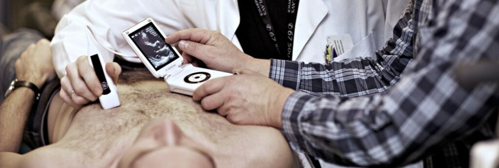
Detecting issues in the foetal heart
How far can we get in detecting problems in the heart or vessels of the foetus during pregnancy? Using blood speckle tracking CIUS aims to detect problems in the heart and blood vessels of the unborn child with CIUS’ new partner, Austrian GE Healthcare Women’s Health Ultrasound (GEWHUS).
Intuitive visualisation
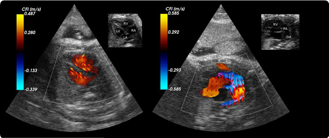
The work on flow estimation and visualisation by the ultrasound group at NTNU led to the launch of the BSI (blood speckle imaging) application in the GE Vivid E95 in 2017.
“We can visualise the blood flow patterns in the heart chambers in a more intuitive way using blood speckle tracking. We are not only estimating the velocity component along the ultrasound beam as we do in the colour Doppler mode, but we can get the 2D velocity estimates and visualise the flow streamlines, which is much more intuitive and accurate,” says Solveig Fadnes, Researcher at NTNU, CIUS.
As of now, this method is available for probes used for cardiac imaging of children. “The anatomy of the heart and the big vessels is quite complex, so if there’s a defect or something is wrong, a more intuitive flow visualisation can help both clinicians and parents understand more,” says Fadnes.
Proceeding from neonatal to foetal diagnosis
The team, consisting of people at CIUS and GEWHUS, aims to develop the BSI application for foetal imaging.
The first step is to get the BSI application incorporated into the Voluson scanner, which is GEWHUS ultrasound scanner for women’s health. “First of all, we want to transfer our knowledge using the ultrasound probes we are using for paediatric patients. We need a purely technical development getting these acquisitions on another type of probe optimising all the technical details,” Fadnes explains.
One essential aim of the project is to find out what they can see when the heart is so small. “It would be very good if we could help the clinicians understand the anatomy and physiology to better predict the outcome for the foetus. This is important in order to provide correct counselling for the parents, especially in cases of severe disease where late abortion is considered,” says Fadnes.
The project is still in the starting block, so it is not yet certain what will be possible. If there’s something wrong with an unborn baby’s heart, it is important to detect it before birth, as some patients need surgery or intervention soon after birth. In Norway, this treatment is only performed at OUS Rikshospitalet in Oslo, and the pregnant mother needs to be transported there before birth. Fadnes points out that several diseases don’t affect foetal life but will become critical after birth, since foetal circulation is very different from that of a delivered baby.
Hoping to learn more about the placenta
Imaging of the placenta might be another project. Placenta issues can cause growth restrictions to the foetus but scanning the placenta itself is not part of the ultrasound examinations of pregnant women in Norway today.
“We want to develop acquisitions so that we can better study the placenta. I think we’ll have data and images to analyse but it’s very difficult to know if it will have a clinical impact. Still, learning more about the physiology of the placenta is also a motivation. Clinicians we have talked to think it’s very interesting,” says Fadnes.
The team
Three people are being funded directly by CIUS in this project, enabled by the partnership with GEWHUS. Along with Solveig Fadnes is Ingvild Kinn Ekroll on the technical side and Karin Deibele starting to work on a clinical PhD in January 2022, with Paediatric Cardiologist Siri Ann Nyrnes as supervisor. There is close collaboration, with regular meetings and updates from each side.
Solveig Fadnes is happy about this collaboration and how it works. She also appreciates the opportunity to work on a clinical high-end scanner: “It is very motivating with a new clinical partner showing interest in what we have been doing and wanting to implement our methods in their scanner.”
This article first appeared in the 2021 CIUS Annual Report.