
Ultrasound and AI improve salmon breeding and welfare
Ultrasound combined with machine learning provides a new, smart, and more reliable way of estimating the maturation states of salmon with the aim of predicting the optimal timing for egg harvesting.
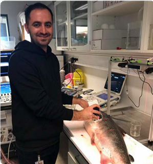
Ultrasound applied to fish is a new venture in CIUS. Yasin Yari is doing his PhD on ultrasound and machine learning for smart monitoring of maturation states in Atlantic salmon in collaboration with AquaGen, Mowi, and CIUS partner InPhase Solutions. His work builds on that of Dr. Ingun Næve (AquaGen), who developed several methods to screen Atlantic salmon maturation states using state- of-the-art medical ultrasound equipment.
Estimating the weight of the ovary
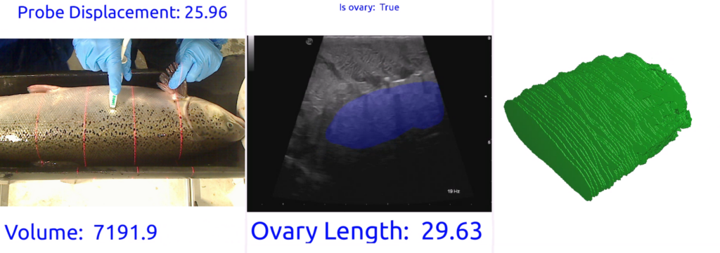
The first task of Yari’s PhD project is to measure the length and volume of the fish ovary and estimate its maturation state. The size gives an indication of the weight.
“We need to know the weight of the ovary in the fish. What we can do now is to use ultrasound to get a very good estimation of the weight of the ovary, and having that, we can predict when the fish will be ready for stripping,” says Yari. Stripping is the removal of the eggs for further farming.
Current methods use an ultrasound probe along with a ruler to measure the length of the ovary manually, but Yari is combining machine learning and ultrasound to automate and improve the accuracy of these methods.
“We are developing a method that can recognise the ovary, measure its length and volume, and estimate the relative weight of the ovary to the fish weight,” says Yari.
To use the automatic maturation estimation method more efficiently, there is a need for the automation of ultrasound image acquisition. So, the long-term plan is to scan the fish whilst in water.
“Scanning underwater removes both the need to take fish into the air and the stress associated with the scanning, which benefits fish health and welfare. Moreover, it simplifies the work and reduces the workload for the staff in the production department,” Yari points out.
Monitoring the development of the eggs
The second task in the PhD project involves smart screening of the development of the eggs. Using ultrasound scanning, it is possible to watch as a dark spot appears in the egg, grows bigger and, in the end, almost fully covers the egg. The fish is ready for stripping at this stage, and the belly will now become soft as the fish ovulates.
“At the moment, the procedure is to take the fish out of the tank and check the belly by palpation. The fish is ready for stripping if the belly is soft, meaning the eggs are released. This must be done for each individual,” says Yari.
He is optimistic that they can find a way of using the ultrasound image to see if the eggs have been released, avoiding the need for the demanding palpation procedure.
Ultrasound also enables them to see if there is a problem in the ovary. If there is blood in the ovary or eggs are not maturing as expected, the ultrasound image will reveal this, and the fish can be removed from the cage.
Measuring the fat content
Finally, the third task is to monitor the fat content of the muscle using ultrasound image analysis.
“The fat content of the fish determines whether the fish will go into the maturation process in that year or if it needs to wait another year to grow and produce more fat before entering the process. The muscle and deep-lying abdominal (visceral) fat content before maturation could perhaps say something about which individuals are more likely to mature, and it can be a selection criterion for broodfish candidates. As maturation progresses, energy stored as fat in muscles and viscera is used in the building of the eggs,” explains Yari.
One challenge that Yari faces is the ultrasound probes, which are not customised for this task. Although common standard transducers are good enough to achieve reliable information, the ideal would be to have an ultrasound transducer that is customised for fish scanning.
Yasin Yari started his PhD in December 2020 and is set to complete it towards the end of December 2023. His super- visors are Professor Lasse Løvstakken at CIUS, Dr. Ingun Næve from AquaGen, Dr. Marco Marien Voormolen from InPhase Solutions, together with Dr. Per Helge Bergtun, who is a veterinarian from Mowi and Dr. Svein-Erik Måsøy who is the Director of CIUS.
This article first appeared in the 2021 CIUS Annual Report.

