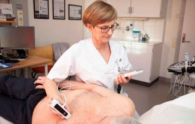
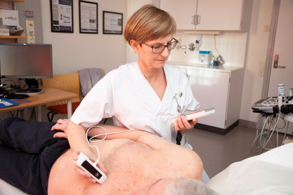
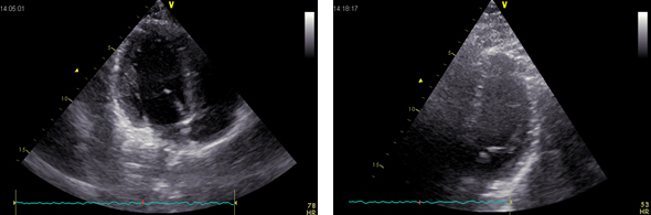
Can AI on hand-held ultrasound help diagnose heart failure?
Artificial intelligence software and telemedical transfer of images have shown promising results in aiding users to evaluate heart function, but in our study the artificial intelligence software on hand-held ultrasound devices alone is not reliable enough to evaluate heart function for general practitioners.
Uncovering stiff or leaky heart valves
Modern ultrasound scanners combined with technological developments at NTNU may improve diagnostics for dis- eases related to narrow or leaky heart valves. Advanced 3D Doppler and high frame rate imaging makes it possible to visualise cardiac flow and minor movements of the heart’s muscular tissue more precisely, according to PhD Candidates Torvald Espeland and Erik Andreas Rye Berg.
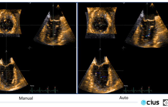
Automating heart monitoring
Heart disease is a major public health issue, and every year, more than 100,000 heart valve operations are performed in the United States alone. With these kinds of numbers, it is more and more urgent to simplify diagnosis and monitoring of heart patients. I/we believe automating the interpretation of ultrasound results is the way forward.
Taking heart imaging out of the hospital with AI
You have certainly heard about Artificial Intelligence (AI), but have you ever wondered what it is used for? If not, an excellent example of Artificial Intelligence is self-driving cars. The car computer acts as a brain, and cameras as eyes. But this is just a…
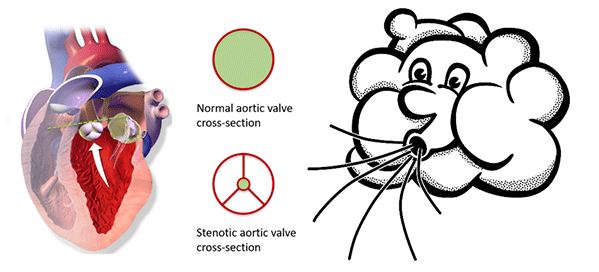
Assessing aortic stenosis severity by ultrasound
Aortic valve stenosis is a narrowing of the valve that separates the left ventricle from the aorta. A reduced opening increases the effort required by the left ventricle to pump blood. Being a degenerative disease, patients with aortic stenosis must undergo a clinical follow-up, which is usually performed by ultrasound. At the Centre of Innovative Ultrasound Solutions (CIUS), we are developing a new method that exploits 3-D high frame-rate imaging to increase the degree of automation in aortic stenosis flow measurements. The goal is to speed up the workflow in the clinics and increase the accuracy of measurements.
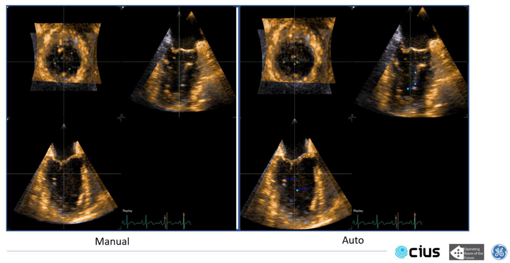
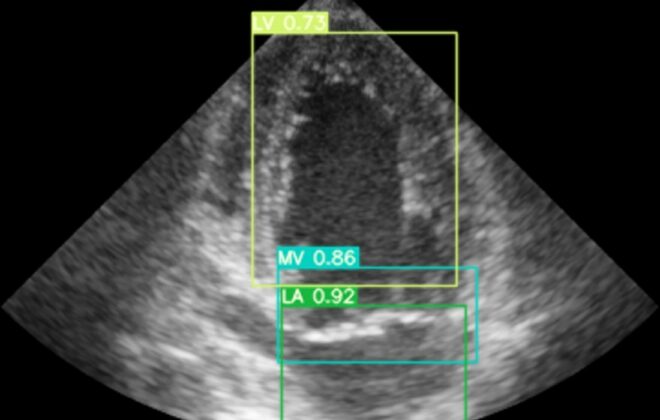
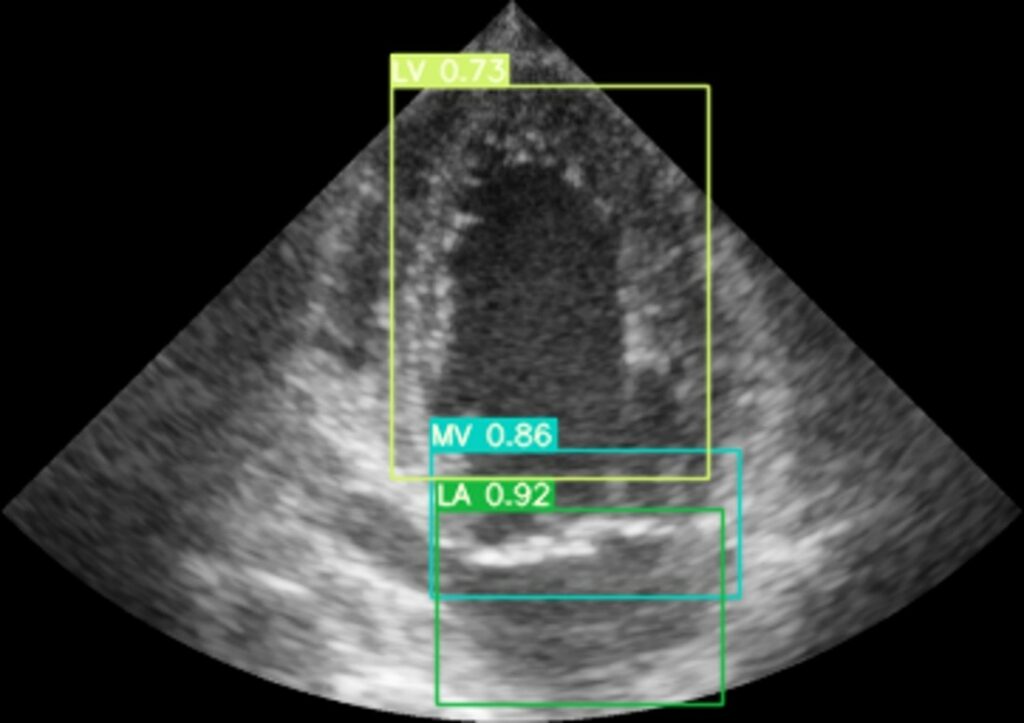
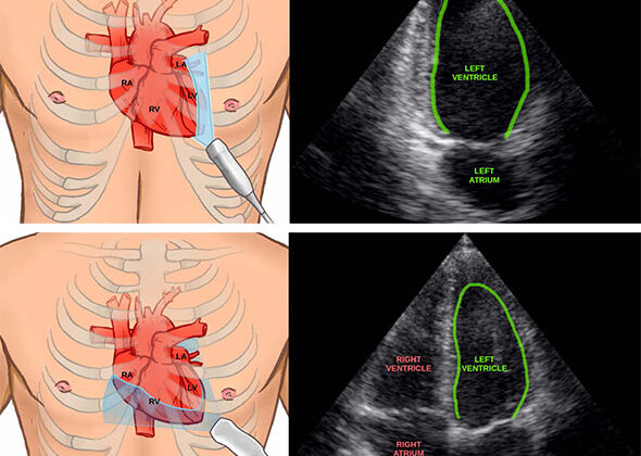
Using artificial intelligence to measure the heart
Artificial intelligence can now help clinicians by automatically measuring the heart in ultrasound images. This can save time and may in the future enable inexperienced users to perform accurate measurements of the heart.
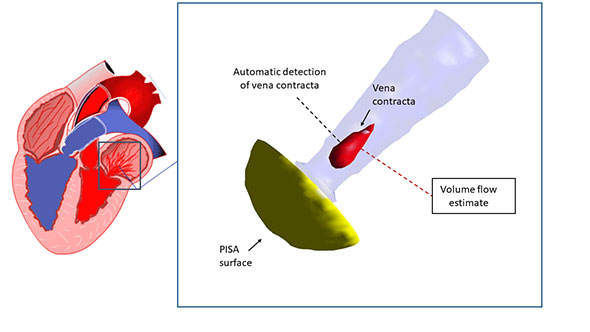
Leaky heart valves – new method for quantification by ultrasound
A leaky heart valve, termed valvular regurgitation, causes some of the blood to flow in the wrong direction. It is one of two major features in valvular heart disease. The patient’s symptoms and prognosis depend heavily on the amount of leakage, which usually is determined…
Pocket-sized ultrasound helps screen stroke patients
When stroke patients are admitted to hospital, the main arteries in the neck are checked for blockages as this is a major cause for blood-clots and stroke. Today, these examinations are done using time-consuming and costly procedures. At NTNU, we have found that using a…
Improving quality of cardiac ultrasound images
Cardiovascular diseases (CVDs) are the number-one cause of death globally. An estimated 17.5 million people died from CVDs in 2012, 31% of all global deaths. One important factor in preventing cardiovascular disease, is early diagnosis using technologies such as ultrasound echocardiography. In this project, we…