
With CIUS at IUS
Twenty-nine experts and researchers associated with CIUS participated at IUS, the IEEE International Ultrasound Symposium in Kobe, Japan, in October 2018. Not only the number of attendees, but also the number of presentations from CIUS was impressively high. The overall scientific contribution from the CIUS…
Mapping early signs of cardiac dysfunction in children using ultrasound
Children with dysfunction of the right heart chamber (ventricle), which pumps blood to the lungs, have lower tolerance to exercise and at risk of sudden cardiac death in more severe cases. This dysfunction usually sets in progressively and detection at earlier stages is crucial to guiding therapies and interventions that improve symptoms and survival. New ultrasound techniques, makes it easier detect and quantify the problem.

Assessing aortic stenosis severity by ultrasound
Aortic valve stenosis is a narrowing of the valve that separates the left ventricle from the aorta. A reduced opening increases the effort required by the left ventricle to pump blood. Being a degenerative disease, patients with aortic stenosis must undergo a clinical follow-up, which is usually performed by ultrasound. At the Centre of Innovative Ultrasound Solutions (CIUS), we are developing a new method that exploits 3-D high frame-rate imaging to increase the degree of automation in aortic stenosis flow measurements. The goal is to speed up the workflow in the clinics and increase the accuracy of measurements.

The health of petroleum wells
How do you check that a petroleum well is leak-proof, so that it cannot endanger the environment or the platform staff? To do so, you’d have to investigate a narrow hole with a diameter of maybe 30 cm, kilometers below the ground, where the temperatures can reach well above 100°C and the pressure is crushing. This may sound difficult, and it is! Even so, the petroleum industry has been doing things like these for almost a century, and over the past four decades they have increasingly been using ultrasonic techniques.
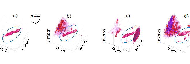
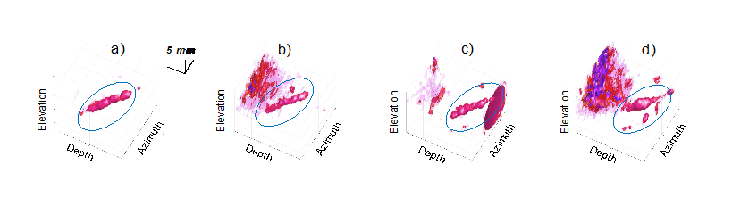
Improving cardiac ultrasound in difficult-to-image patients
Despite constant improvements within the field of medical ultrasound, there are still a considerable number of difficult-to-image patient. Echocardiograms (heart images) taken from these patients do not have the quality that is needed for correct diagnosis. Therefore it is important to further improve the quality of echocardiograms.
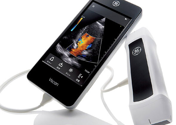
Could your local doctor diagnose heart disease using a handheld ultrasound device?
Many potential heart patients that are referred to specialists, turn out not to need specialist care. If general practitioners (GPs) could use handheld ultrasound devices with built-in diagnostic tools, could this improve patient outcome and reduce cost for the health services?
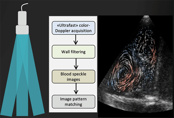
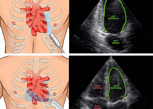
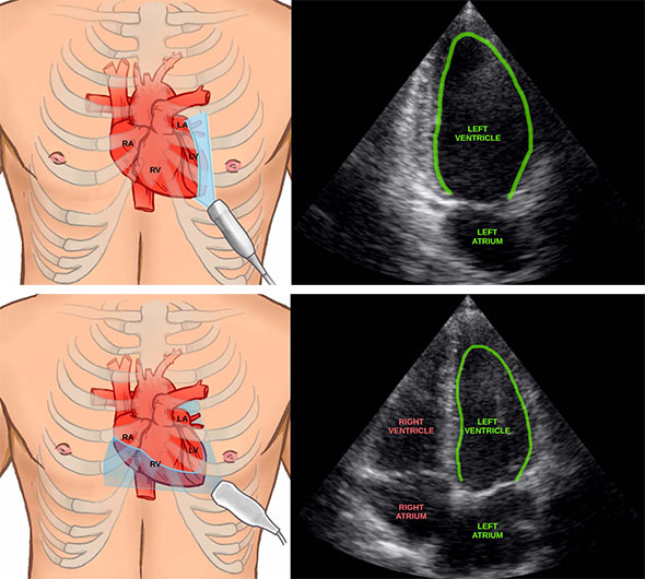
Improving ultrasound images of the heart’s blood vessels
Coronary heart disease is a condition where the heart muscle (myocardium) does not receive enough oxygen and nutrients due to obstruction of blood flow in the heart’s blood vessels, known as the coronary arteries. It is therefore important to investigate the blood flow in these vessels. However, it is challenging to obtain accurate measurements due to the combination of poor blood flow, the constant movement of the heart throughout the cardiac cycle, and surrounding tissue causing noise signals.
Using artificial intelligence to measure the heart
Artificial intelligence can now help clinicians by automatically measuring the heart in ultrasound images. This can save time and may in the future enable inexperienced users to perform accurate measurements of the heart.

Making analog-to-digital converters for digital ultrasound probes
Making good digital ultrasound probes is extremely challenging because of the constrained amount of power that can be used. If significantly more than a couple of watts are consumed, the probe gets too hot to be allowed to touch your skin.
DeepEcho: Machine learning for improved echocardiography
Increased availability of ultrasound devices is great news, but it also triggers a range of new challenges. At CIUS we believe modern machine learning methods, such as deep learning, can provide necessary solutions.